
Cortex Cannibal / Brain & Behaviour
What Alcohol Does to Your Brain Might Be Stranger Than We Thought
New research suggests the brain's immune cells and the molecular structures surrounding neurons may contribute to some effects of heavy alcohol exposure.

Have you ever watched someone who's had far too much to drink insist they're perfectly fine, while their legs seem to have an entirely different opinion?
Their words get slower, their balance becomes questionable, and suddenly walking in a straight line requires a level of concentration normally reserved for defusing a bomb.
We've known for decades that alcohol interferes with how neurons communicate. That's part of why judgment, coordination, memory, and alertness can all go downhill after drinking.
But what if some of those effects involve more than neurons?
A 2026 study published in Immunity suggests the brain's resident immune cells may contribute to alcohol-related sedation and motor impairment by disrupting the molecular environment around neurons. The causal evidence comes from experiments in mice. [1]
And the researchers did something particularly interesting to test it.
The Short Answer
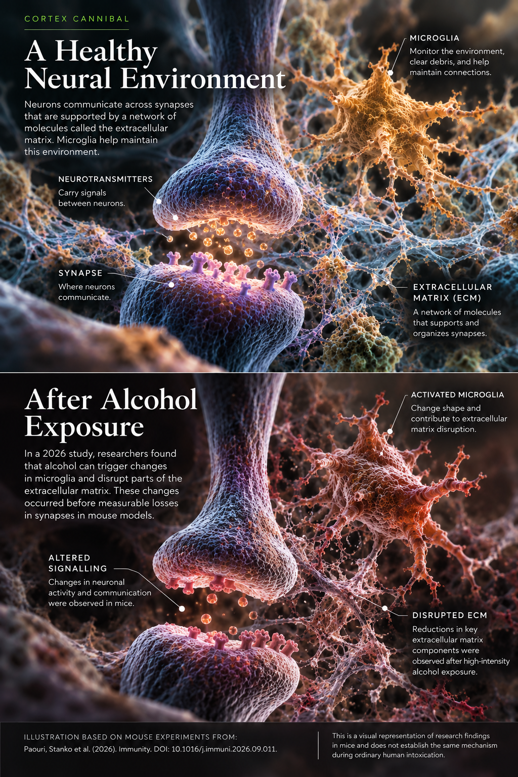
In mice, alcohol exposure prompted changes in microglia, the brain's resident immune cells. Those cells contributed to disruption of the extracellular matrix, a network of molecules that helps support neuronal connections. Genetically removing a microglial signalling protein called MyD88 reduced several alcohol-related impairments. Whether this same mechanism drives intoxication in people remains unknown. [1]
Meet the Brain's Maintenance Crew
Most of us picture the brain as billions of neurons sending electrical and chemical messages back and forth. But there's an entire supporting cast keeping that system running.
Microglia are part of it. These immune cells monitor their surroundings, help clear cellular debris, and participate in maintaining and remodeling connections between neurons.
They're essentially a maintenance crew that never clocks out.
Usually, that work is essential. Microglia aren't inherently harmful, and an immune response isn't automatically a problem. But particular responses, under particular conditions, can contribute to damage.
In this study, researchers exposed mice to alcohol and used imaging to follow microglia in living brain tissue. The cells changed shape before the animals became sedated, and responses became more pronounced with repeated exposure. [1, Figure 1]
That raised an interesting possibility: perhaps microglia weren't simply reacting to an impaired brain. Perhaps they were contributing to some of the impairment.

The Invisible Scaffolding Around Your Neurons
Here's something we don't talk about nearly enough: neurons aren't floating around in empty space.
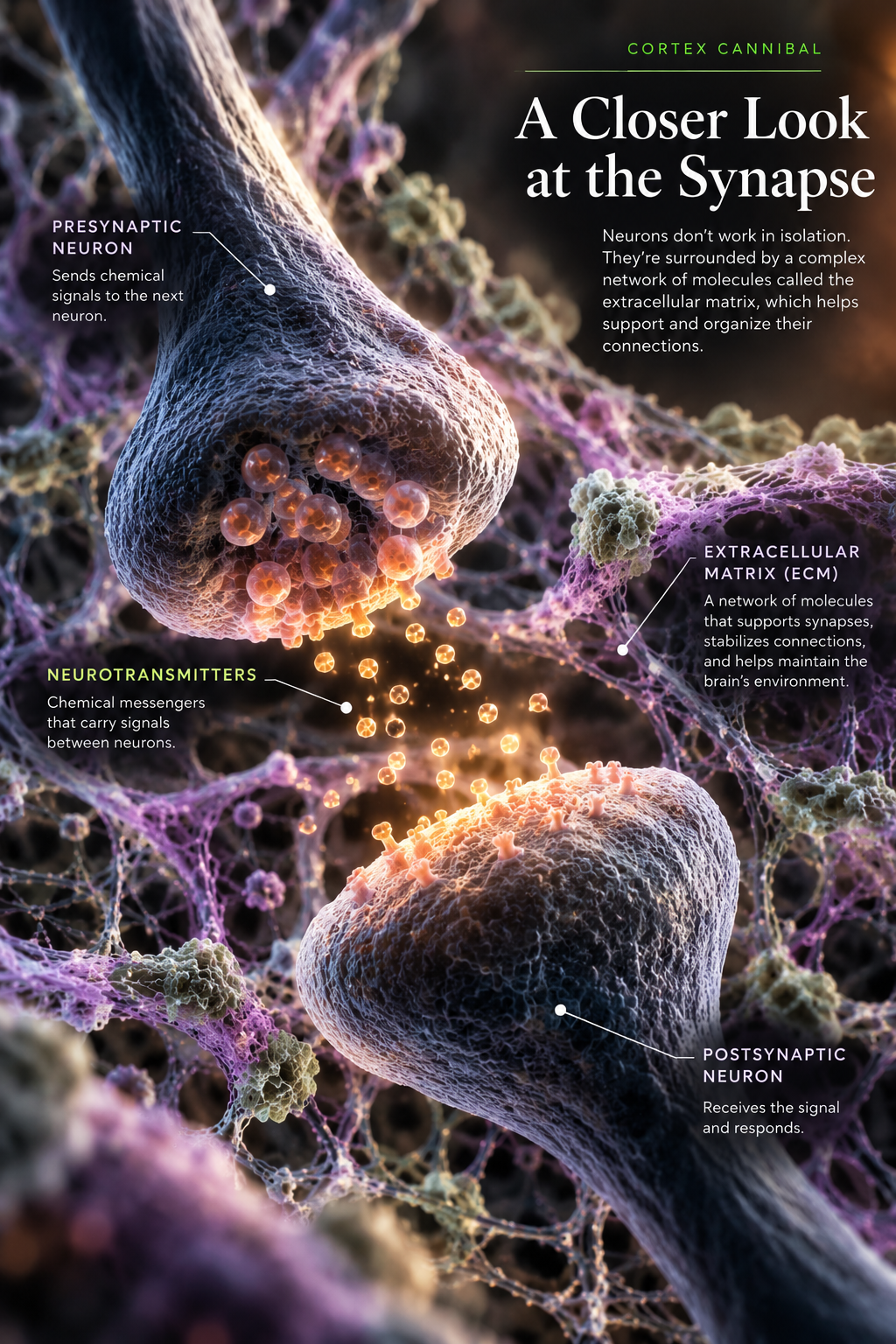
They're surrounded by a network of molecules called the extracellular matrix, or ECM. It helps organize the environment around brain cells and supports the stability and function of their connections.
Think of neurons as traffic moving through a city. The extracellular matrix is part of the infrastructure helping everything stay organized.
In the alcohol-exposed mice, researchers found reductions in several components of this molecular scaffolding. They also observed microglia taking up some of that material. In one exposure model, extracellular matrix changes appeared before measurable synapse loss. [1, Figure 6]
A synapse is the tiny junction where neurons communicate. The extracellular matrix is different: it's part of the environment around those junctions. Damage to one doesn't automatically mean the other has disappeared.
But the findings suggest alcohol may disturb more than the chemical messages travelling between neurons. The structures helping support those connections can change too.
What Happened When Scientists Interrupted the Process?
This is the part of the study I find most interesting.
MyD88 is a protein that helps certain immune signals travel inside cells. Think of it as one part of the communication system that tells a cell how to respond.
The researchers genetically removed MyD88 from microglia in mice. They weren't giving the animals a medication, and they weren't switching off the entire immune system.
After alcohol exposure, these mice showed less extracellular matrix disruption, better preservation of neuronal activity, and less impairment on tests of coordination and sedation than comparison mice. [1, Figures 3, 4 and 6]
But the researchers went further. They used an enzyme called chondroitinase ABC to break down parts of the extracellular matrix directly. When they did, the protection associated with removing microglial MyD88 was reduced or lost, and alcohol-related impairments and synaptic damage increased. [1, Figure 7]
That's important. It helps establish that extracellular matrix disruption was contributing to the effects in these mouse experiments, rather than simply appearing alongside them.
They also compared alcohol with pentobarbital, another sedative. Removing microglial MyD88 did not protect against pentobarbital's sedating effects in the same way. [1, Figure 4]
So this wasn't just a case of making mice generally harder to sedate. It pointed toward a pathway involved in alcohol's effects.

Does This Happen in Human Brains?
Possibly, but we don't have the same level of evidence yet.
The researchers examined frontal cortex tissue from six people with alcohol use disorder and six control cases. They found reductions in two extracellular matrix components, hyaluronan and versican, in the alcohol use disorder samples. [1, Figure 6]
That's an interesting connection to the mouse findings. But these were postmortem samples. They cannot establish when the changes happened, whether microglia caused them, or whether they contributed to intoxication.
There's another important distinction: the mouse experiments involved controlled binge-like and high-intensity alcohol exposure. The human tissue came from people with alcohol use disorder.
We can't use this study to claim that having one drink causes the same changes, or that the mouse mechanism explains an ordinary night of drinking in humans.
The science simply isn't there yet.
Why This Changes the Conversation
For decades, much of our understanding of alcohol's immediate effects has centred on neurons and neurotransmitters. That explanation remains important.
But the brain is more than its neurons.
This research adds another layer: immune signalling and the molecular environment around synapses may contribute to some neurological consequences of heavy alcohol exposure.
It also gives scientists a pathway worth investigating. That doesn't mean a treatment is around the corner. MyD88 has important immune functions, and genetically changing mice is very different from safely treating humans.
Sometimes understanding the brain means paying attention to the cells that don't get nearly as much attention as neurons.
And apparently, even the maintenance crew can get caught up in the chaos.
Still Curious?
Frequently Asked Questions
Does alcohol actually damage brain cells?
Heavy or repeated alcohol exposure can damage the brain. But not every immediate effect of intoxication represents permanent cell damage. This study found changes involving neuronal activity, synapses, and neuronal integrity in mice. It doesn't establish the amount of damage caused by an individual drinking episode in humans.
What are microglia, and why do they matter?
Microglia are resident immune cells in the brain. They monitor their surroundings and help maintain neural tissue. In this study, a particular microglial signalling pathway contributed to extracellular matrix disruption and neurological impairment after alcohol exposure in mice.
Can the brain repair its extracellular matrix?
The extracellular matrix can undergo remodeling. But this study doesn't establish how quickly or completely the alcohol-related changes it observed might recover, especially in people.
Does this mean scientists can prevent drunkenness?
No. Genetic changes reduced some alcohol-related impairments in mice. They do not provide a clinically validated treatment, and alcohol still has direct effects on neuronal signalling.
One Last Thought
There's More Going On Up There Than We Realize
We tend to think of intoxication in terms of what we can see. Someone loses their balance, forgets what they were saying, or becomes convinced they're suddenly an excellent singer.
But underneath all that, the brain is managing an enormous amount of cellular activity.
Neurons communicating. Immune cells responding. Molecular structures being maintained and remodeled.
It's easy to forget that the brain is living tissue, not just a collection of electrical signals.
We've spent decades studying what alcohol does to our neurons, and we're still finding other cells and structures that may be part of the story.
Being human is weird. Stay curious.
Follow the Evidence
Research & References
[1] Paouri, E., Stanko, S., et al. (2026). Microglia-mediated extracellular matrix disruption drives neuronal dysfunction, motor impairments, and sedation following alcohol abuse. Immunity. Read the original study ↗
Specific results discussed above are drawn from Figures 1, 3, 4, 6 and 7 of the primary study.
Evidence note: The causal experiments were performed in mice. The human postmortem tissue comparison does not establish the same causal mechanism during human intoxication.
Visual Disclosure
Some illustrations in this article were created using artificial intelligence for educational and visual purposes. They are not actual images from the referenced research.
Medical & Scientific Disclaimer
Cortex Cannibal publishes educational content about neuroscience, health, and human behaviour. This article discusses research primarily conducted in mice, with limited supporting observations from human brain tissue. It is not medical advice, diagnosis, or treatment. If you have concerns about alcohol use or its effects on your health, consult a qualified healthcare professional.